Laurie's Blogs.
May 2025
Accessoriometacarpal Ligament Injury in (my) Dog

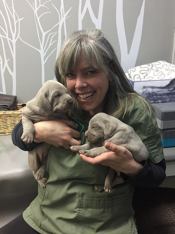
Figure 1: Evidence that he is astonishingly handsome.
FigJam, a 5 year old, devilishly handsome (Figure 1), neutered Labrador retriever and my favourite adventure buddy, developed an intermittent left forelimb lameness that would transiently appear following off leash hikes in the woods. The lameness was mild, lasting only a few strides when he first got up from a rest. Pain could be localized to the left accessory carpal bone (ACB), and there was mild swelling distal to it.
Radiographs showed enthesophytic changes to the distal apical aspect of the ACB (Figure 2). Ultrasound imaging of the area revealed regional loss of echoarchitecture, reduced echogenicity, and mild swelling in the superficial half of the accessoriometacarpal ligament of the 5th digit (Figure 3).

Figure 2: Lateral carpal radiograph showing bony proliferation of the distal apical aspect of the ACB

Figure 3: Ultrasound images of the accessoriometacarpal ligament at the time of diagnosis
Thanks, buddy. That’s a new one.
Apparently, dogs have two accessoriometacarpal ligaments (Figure 4), both of which originate from the distal apical end of the ACB and insert on the proximal palmar metacarpus – one on the 5th metacarpal bone, and one on the 4th. Having never seen this injury before, and unable to find anything in the literature, I treated it as I would any other ligamentous injury. Given the relatively mild presentation and the small size of the lesion, I elected for extracorporeal shockwave therapy (ESWT) instead of stem cells as the primary modality.

Figure 4: Plantar view of the carpus showing the accessoriometacarpal ligament running from the ACB (marked CA in this image) to the proximal 5th metacarpal bone. Image taken from: Brinker, Piermattei, and Flo’s Handbook of Small Animal Orthopedics and Fracture Repair, 5th ed.
Because the lameness only occurred following episodes of off leash running in challenging terrain, FigJam was restricted to leash walks on an extendable lead, on mostly easy terrain, without limiting the duration of the walks. Radial ESWT was administered 4 times (2000 shocks, 10Hz, 2 bars), immediately followed by photobiomodulation (Spectravet 16J at the skin surface, 500mW, 810 nm). These treatments were given over a 13-week period.
After 3 weeks of not seeing any lameness, we both grew a bit itchy to return to more interesting walks. To facilitate this, FigJam was fitted for a neoprene brace (Therapaws) to provide additional carpal support while running. He would only wear the brace during off-leash hikes in the forest, and it was enough to prevent any evidence of stiffness following that level of exercise. He lost the first brace somewhere toward the end of one hike, so I replaced it… but it only took a few weeks for him to lose that one too.
Repeat ultrasound 9 weeks following the initial showed good evidence of healing of the accessoriometacarpal ligament (Figure 5), with infilling of the lost architecture and resolution of the swelling. Hypoechoic changes persisted, but this amount of repair in such a short time was encouraging.

Figure 5: Repeat ultrasound images showing infilling of the affected ligament.
Because FigJam lost the 2nd brace halfway through the hike, and showed no signs of stiffness afterward, he was returned to 1 hour off leash hikes in the woods, progressing to 2 hours, and from moderately challenging (blue square) to very challenging (black diamond) terrain, and then back to full activity. It has been a just shy of a year since then, and the lameness has not recurred.
I have since seen 1 other reference to this condition as part of a lecture on unusual carpal injuries, so apparently FigJam isn’t the only one to get this. He doesn’t always show perfect wisdom when selecting what rock features to jump off of, which I can see as being an acute cause of this injury. However, there were some radiographic changes to his other ACB as well, suggesting more of a repetitive stress insult… or some combination of the two.
Either way, he is back to full function with all his enchantingly good looks unaffected.
About the Author:
Dr. David Lane owns and operates Points East West Veterinary Services based out of Squamish, BC (Canada). https://www.pointseastwest.com/ He is a specialist in canine sports medicine and rehabilitation therapy. His research on the clinical effectiveness of combining chiropractic techniques with acupuncture therapy in dogs is the first of its kind. He was the first researcher to demonstrate the link between lower back pain and canine urinary incontinence; that treating back pain can reduce or eliminate symptoms in the majority of dogs. He also developed and published a minimally invasive surgical technique for resolving biceps tendon injury in dogs.
Current areas of interest include documenting success rates when using stem cells to repair partially torn tendons, and developing a treatment algorithm for addressing injury to the biceps tendon.
His work with sporting dogs has brought him to numerous regional and national events, as well as travelling with the national team to the World Agility Championships in Spain and the Netherlands.
PS He's looking for another rehab vet to come work with him. Check out his practice!!