Laurie's Blogs.

Nov 2025
Addressing Neurogenic Muscle Atrophy in Canines: Why Exercise Alone Isn’t Enough
In canine rehabilitation, muscle atrophy is a common challenge, particularly when it’s neurogenic—caused by neuritis of spinal origin. Veterinary professionals, physiotherapists, or canine conditioning coaches may encounter clients eager to restore their dog’s muscle mass through exercise programs, but when atrophy stems from nerve dysfunction, such efforts can be futile unless the underlying nerve pathology is addressed. This is especially true when neuritis results from a pinched nerve due to mechanical joint dysfunction in the canine spine or sacroiliac joint. In this blog, we’ll explore why targeting the root cause is critical for effective rehabilitation, define key terms, and provide guidance for canine health professionals or those working in canine conditioning practice.
Defining Key Terms for Canine Rehabilitation
To understand the limitations of exercise in treating neurogenic atrophy in dogs, let’s clarify some essential terms:
• Neuritis: This is inflammation of a peripheral nerve, which in dogs may manifest as lameness, weakness, pain, or reduced mobility. In people, it often causing symptoms like pain, numbness, tingling, or weakness in the affected area. Inflamed nerves struggle to transmit signals to muscles, leading to impaired function. It might also be known as neurapraxia.
• Neurogenic Atrophy: This occurs when muscle wasting results from compromised nerve function, disrupting the nerve signals required for muscle activation and maintenance. Unlike disuse atrophy (e.g., from immobilization after surgery), neurogenic atrophy is directly linked to nerve pathology, such as spinal nerve root issues.
• Nerve Root Impingement: Also called radiculopathy, this involves compression or irritation of a spinal nerve root, often due to mechanical issues like a lateralized intervertebral disc lesion, disc degeneration, vertebral malalignment, or sacroiliac joint dysfunction. This compression (or sometimes ‘stretch’) of the nerve disrupts nerve signals, causing weakness or atrophy in the muscles innervated by the affected nerve.
Why Exercise Alone Falls Short for Neurogenic Atrophy
When a dog presents with muscle atrophy due to neuritis of spinal origin, the problem lies in the nervous system, not the muscle itself. The affected muscles waste away because they’re not receiving adequate nerve signals to contract or maintain mass. While exercise is a cornerstone of canine rehabilitation for many conditions, targeting neurogenic atrophy with strengthening exercises—such as controlled walking, hydrotherapy, or specific exercises—won’t yield results if the nerve signal remains compromised. In fact, overexertion could worsen inflammation or discomfort, potentially delaying recovery.
Consider a dog with hindlimb atrophy due to a pinched L4, L5, or L6 nerve root from disc degeneration, a common issue in large breeds like Retrievers, Shepherds, or Rottweilers. Exercises like sit-to-stand repetitions or underwater treadmill therapy may seem like logical interventions, but if the nerve is compressed or inflamed, the muscles (e.g., quadriceps, hamstrings, or adductors) won’t respond effectively. The focus must shift to resolving the nerve pathology before expecting exercise to rebuild muscle.
The Role of Spinal and Sacroiliac Joint Dysfunction in Canines
Neuritis leading to neurogenic atrophy in dogs is often linked to mechanical joint dysfunction in the spine or sacroiliac joint. A “pinched nerve” in these cases typically results from:
• Spinal Joint Dysfunction: Conditions like IVDD (lateralized disc or a disc protrusioin at the cauda equina), vertebral instability, or intervertebral disc degeneration can compress nerve roots, causing inflammation and impairing nerve function. For example, a degenerative disc in the lumbar spine may compress the L6 nerve root, leading to atrophy in the stifle extensors or hip adductors.
• Sacroiliac Joint Dysfunction: Misalignment or inflammation in the sacroiliac joint can irritate nerves like the sciatic nerve, contributing to muscle weakness or atrophy in the hindlimbs, such as the gluteals or hamstrings. Poor alignment of functioning of the sacroiliac joints might also compromise the function or position of the spinal segments of the caudal lumbar spine as well, thus leading to lumbar spine neuritis.
In both scenarios, the nerve root impingement or inflammation disrupts neural input to the muscle, causing atrophy. Exercise programs aimed at strengthening the affected muscles won’t restore function if the nerve signal is blocked. Veterinary professionals must prioritize relieving the nerve compression or inflammation to halt atrophy and enable effective rehabilitation.
A Veterinary Rehabilitation Approach
For canine physiotherapists and veterinary professionals, managing neurogenic atrophy requires a focus on diagnosing and treating the underlying nerve pathology. Here’s a practical approach:
1. Accurate Diagnosis: There are two ways to approach this. My favourite being a physical examination by someone with the skills to evaluate for a spinal, or sacroiliac joint dysfunction. Then a course of treatment to attempt to alleviate the symptoms. From my experience this is the most practical and cost effective first step. Alternately, beginning with imaging (e.g., MRI, CT) and a neurological exam to confirm nerve root impingement or neuritis might identify the cause, such as IVDD. Electromyography (EMG) can help assess nerve and muscle function. However expensive diagnostics might not reveal the root of the neural dysfunction (e.g. in the case of a sacroiliac joint dysfunction) and are not likely to change the primary treatment plan. They might be better left as a secondary option, should a physical exam and conservative management fail to resolve the issue.
2. Targeted Interventions: Depending on the severity, treatments may include:
• Manual Therapy: Veterinary chiropractic adjustments or physiotherapy techniques to correct spinal or sacroiliac joint dysfunction and relieve nerve compression.
• Medical Management: Anti-inflammatory medications (NSAIDS) may be in order to reduce neuritis and swelling around the nerve
• Supplements: Some owners might seek nutraceutical options that are purposed to be effective for inflammation.
• Modalities: Therapeutic modalities such as photobiomodulation (laser) or pulsed electromagnetic field therapy might have a role in conjunction with manual therapies and NSAIDS or nutraceuticals.
• Surgical Options: For severe cases, such as advanced IVDD causing significant nerve compression, surgical decompression (e.g., hemilaminectomy) may be necessary. Personally, I think this is only an options once all conservative measures have been exhausted.
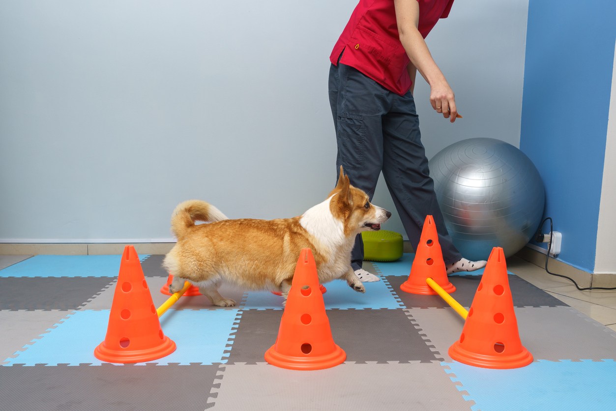
3. Rehabilitation with Exercise: Once nerve function begins to improve, tailored exercise programs can help rebuild muscle strength and prevent further atrophy. Hydrotherapy or controlled exercises (e.g., use of cavaletti poles, inflatable equipment, or sit to stand exercises) should be introduced gradually and customized to the dog’s recovery stage.
Summary: Treat the Nerve, Then the Muscle
For veterinary rehabilitation professionals working with canine patients, the key to addressing neurogenic muscle atrophy caused by neuritis of spinal origin is clear: exercise alone is ineffective if the underlying nerve pathology remains unresolved. Whether the neuritis stems from a pinched nerve due to IVDD or sacroiliac joint dysfunction, the priority is to relieve nerve compression or inflammation. Only then can therapeutic exercises effectively restore muscle function and improve mobility. By focusing on the root cause first, veterinary professionals can guide dogs toward a full and lasting recovery.