Laurie's Blogs.

Nov 2026
Percutaneous Laser Disk Ablation in Dogs
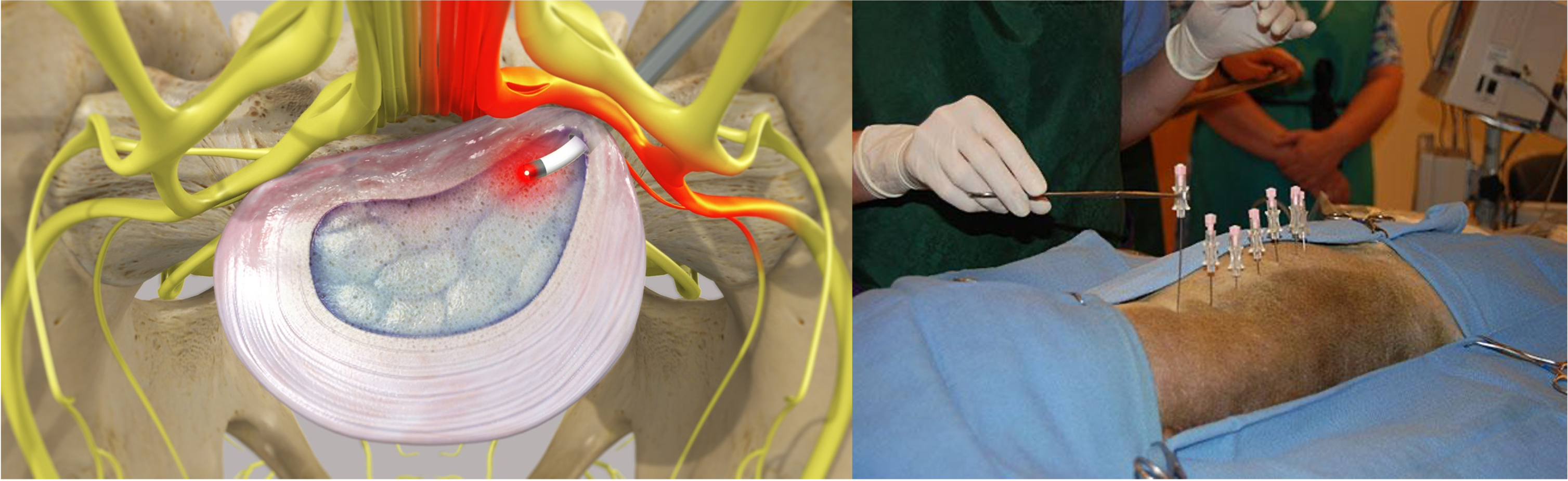
Have you heard of this? Have you seen a dog that has received this treatment? I’ve not, but it sounds interesting!! Percutaneous laser disk ablation (PLDA), primarily using a holmium:YAG (Ho:YAG) laser, is a minimally invasive prophylactic procedure developed mainly at Oklahoma State University to reduce the risk of recurrent thoracolumbar intervertebral disk herniation (IVDH) in dogs (especially chondrodystrophic breeds) that have recovered from a prior episode (Bartels et al., 2003; Dugat et al., 2016).
Key Clinical Studies (Ho:YAG PLDA)
- **Foundational 1996 study**: In 33 dogs with recurrent back pain after conservative management of disk disease, percutaneous needles were placed under fluoroscopy into the nucleus pulposus of disks T10-11 through L3-4. Ho:YAG laser energy ablated the disk contents. All dogs recovered without complications. Radiography and histology showed reduced nucleus pulposus volume. The technique was proposed for both treatment of partially herniated disks and prophylaxis against future herniations (Dickey et al., 1996).
- **2003 outcome study**: Retrospective review of 277 dogs (1992–2001) in which the same 7 thoracolumbar disks were ablated prophylactically. Of 262 dogs with follow-up (mean ~15 months), only 9 (3.4%) had recurrence of paresis/paralysis (mean 15 months post-procedure). Acute complications were uncommon (mild pneumothorax, needle-site abscess, transient proprioceptive deficits in a few dogs; one required hemilaminectomy; one later diskospondylitis). The authors concluded it had few complications and appeared to lower recurrence risk (Bartels et al., 2003).
- **2016 recurrence study**: 303 dogs (1994–2011) with prior suspected/confirmed thoracolumbar IVDH treated by PLDA and followed ≥3 years (or until documented recurrence). 60/303 (19.8%) had a suspected/confirmed subsequent episode, but only 11/303 (3.6%) had imaging- and surgery-confirmed recurrence. Recurrence rates did not differ significantly between dogs previously managed medically vs. surgically. Owner satisfaction was high (≥9/10 in ~93%; most dogs same or improved immediately post-procedure). The procedure was described as relatively safe and associated with low confirmed recurrence (Dugat et al., 2016).
- **2021–2022 MRI evaluation**: Assessed perioperative changes to disks, vertebral endplates, and spinal cord after thoracolumbar PLDA. Mild diskitis and endplate lesions were commonly visible on MRI in treated disks, with some increased disk degeneration scores at certain levels. No disk extrusions or spinal cord lesions were identified, and changes did not correlate with clinical complications. The authors concluded PLDA remains a safe, effective option to reduce IVDH recurrence despite these imaging findings (Irizarry et al., 2022).
Supporting / Related Research
- Early animal work (1994 Chinese study) used Nd:YAG laser (1.06 µm) via a transperitoneal approach to ablate lumbar disks in 12 dogs; fibrous tissue later proliferated, with limited cartilage regeneration (Qi et al., 1994).
- A 2009 study evaluated 980-nm diode and 1064-nm Nd:YAG lasers (in vitro on bovine disks + in vivo canine model). It standardized parameters and recommended Nd:YAG (initial ~40 W, then 10–15 W; total energy density ~1500–2000 J/cm² with bare fiber) for clinical use based on ablation depth and thermal effects (Jayasree et al., 2009).
- Related 2014 work explored percutaneous single-fiber reflectance spectroscopy as a potential in-situ tool (compatible with PLDA needle placement) to assess mineral degeneration in canine disks, which could allow energy dose adjustment; it showed some correlation with scattering changes from mineralization but limited diagnostic performance versus CT/radiography (Piao et al., 2014).
**Overall consensus from the literature**: PLDA (typically Ho:YAG, targeting T10–L4 or similar thoracolumbar disks) is a low-morbidity prophylactic option for dogs with prior IVDH that have recovered. Confirmed recurrence rates in large series are low (~3–4%), complications are infrequent, and owner satisfaction is high. Imaging changes (endplate lesions, mild diskitis) occur but are usually clinically silent. It is not a substitute for decompression in acute severe cases and is mainly performed at specialized centers (notably Oklahoma State) (Bartels et al., 2003; Dugat et al., 2016; Irizarry et al., 2022). Other lasers (Nd:YAG, diode) have been studied experimentally but Ho:YAG is the primary clinical modality in dogs (Dickey et al., 1996; Jayasree et al., 2009).
References
1.Bartels, K. E., Higbee, R. G., Bahr, R. J., Galloway, D. S., Healey, T. S., & Arnold, C. (2003). Outcome of and complications associated with prophylactic percutaneous laser disk ablation in dogs with thoracolumbar disk disease: 277 cases (1992–2001). Journal of the American Veterinary Medical Association, 222(12), 1733–1739. https://doi.org/10.2460/javma.2003.222.1733
2.Dickey, D. T., Bartels, K. E., Henry, G. A., Stair, E. L., Schafer, S. A., Fry, T. R., & Nordquist, R. E. (1996). Use of the holmium yttrium aluminum garnet laser for percutaneous thoracolumbar intervertebral disk ablation in dogs. Journal of the American Veterinary Medical Association, 208(8), 1263–1267. https://doi.org/10.2460/javma.1996.208.08.1263
3.Dugat, D. R., Bartels, K. E., & Payton, M. E. (2016). Recurrence of disk herniation following percutaneous laser disk ablation in dogs with a history of thoracolumbar intervertebral disk herniation: 303 cases (1994–2011). Journal of the American Veterinary Medical Association, 249(12), 1393–1400. https://doi.org/10.2460/javma.249.12.1393
4.Irizarry, I. N., Dugat, D. R., Sippel, K. M., & Payton, M. E. (2022). Evaluation of the intervertebral disk, vertebral body, and spinal cord for changes secondary to percutaneous laser disk ablation. Veterinary Surgery, 51(1), 97–108. https://doi.org/10.1111/vsu.13684
5.Jayasree, R. S., Gupta, A. K., Bodhey, N. K., & Mohanty, M. (2009). Effect of 980-nm diode laser and 1064-nm Nd:YAG laser on the intervertebral disc—In vitro and in vivo studies. Photomedicine and Laser Surgery, 27(4), 547–552. https://doi.org/10.1089/pho.2008.2304
6.Piao, D., McKeirnan, K. L., Sultana, N., Breshears, M. A., Zhang, A., & Bartels, K. E. (2014). Percutaneous single-fiber reflectance spectroscopy of canine intervertebral disc: Is there a potential for in situ probing of mineral degeneration? Lasers in Surgery and Medicine, 46(6), 508–519. https://doi.org/10.1002/lsm.22261
7.Qi, Q., Dang, G. D., & Cai, Q. L. (1994). Laserablation of intervertebral disc: Animal experiment. Zhonghua Wai Ke Za Zhi [Chinese Journal of Surgery], 32(3), 187–189.